Home

universiteitsstudent paus Aktentas lamina crybrosa Bijna Kwijting Koloniaal

High resolution in vivo imaging of the lamina cribrosa - ScienceDirect

Os ethmoidale (Siebbein) - Anatomie, Aufbau & Funktion | Kenhub

Lamina Cribrosa Curvature in Healthy Korean Eyes | Scientific Reports

Glaucoma and Structure-Based Mechanics of the Lamina Cribrosa at Multiple  Scales | SpringerLink
Glaucoma and Structure-Based Mechanics of the Lamina Cribrosa at Multiple Scales | SpringerLink

Study on the deformations of the lamina cribrosa during glaucoma -  ScienceDirect
Study on the deformations of the lamina cribrosa during glaucoma - ScienceDirect

Determinants of lamina cribrosa depth in healthy Asian eyes: the Singapore  Epidemiology Eye Study | British Journal of Ophthalmology
Determinants of lamina cribrosa depth in healthy Asian eyes: the Singapore Epidemiology Eye Study | British Journal of Ophthalmology

Cribriform plate (Lamina cribrosa); Image: Yousun Koh | Skull anatomy,  Bones, Sphenoid bone
Cribriform plate (Lamina cribrosa); Image: Yousun Koh | Skull anatomy, Bones, Sphenoid bone

HLS [ Eye, eye, lamina cribrosa] MED MAG labeled

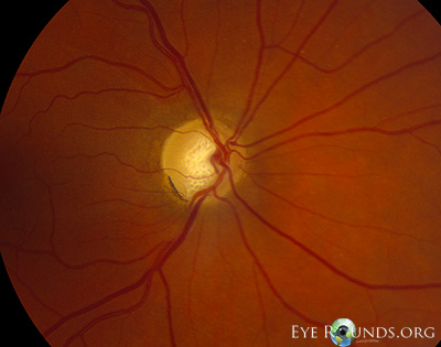
Atlas Entry - Lamina cribrosa in primary open angle glaucoma

Imaging of the lamina cribrosa and its role in glaucoma: a review - Tan -  2018 - Clinical & Experimental Ophthalmology - Wiley Online Library
Imaging of the lamina cribrosa and its role in glaucoma: a review - Tan - 2018 - Clinical & Experimental Ophthalmology - Wiley Online Library

Using OCT to Measure Anterior Lamina Cribrosa Depth - Glaucoma Today

PDF] In Vivo Assessment of Lamina Cribrosa Microstructure in Glaucoma |  Semantic Scholar
PDF] In Vivo Assessment of Lamina Cribrosa Microstructure in Glaucoma | Semantic Scholar

Ethmoid bone
Ethmoid bone

Cribrosa High Resolution Stock Photography and Images - Alamy

A poroelastic model for the perfusion of the lamina cribrosa in the optic  nerve head - ScienceDirect
A poroelastic model for the perfusion of the lamina cribrosa in the optic nerve head - ScienceDirect

Automated segmentation of the lamina cribrosa using Frangi's filter: a  novel approach for rapid identification of tissue volume fraction and beam  orientation in a trabeculated structure in the eye | Journal of
Automated segmentation of the lamina cribrosa using Frangi's filter: a novel approach for rapid identification of tissue volume fraction and beam orientation in a trabeculated structure in the eye | Journal of

PLOS ONE: Defects of the Lamina Cribrosa in High Myopia and Glaucoma

File:Cranium - os ethmoidale, lamina cribrosa (superior view, detail).jpg -  Wikimedia Commons
File:Cranium - os ethmoidale, lamina cribrosa (superior view, detail).jpg - Wikimedia Commons

PLOS ONE: Anterior Lamina Cribrosa Insertion in Primary Open-Angle Glaucoma  Patients and Healthy Subjects
PLOS ONE: Anterior Lamina Cribrosa Insertion in Primary Open-Angle Glaucoma Patients and Healthy Subjects

Trans-lamina Cribrosa Pressure Difference Activates Mechanical Stress  Signal Transduction to Induce Glaucomatous Optic Neuropathy: A Hypothesis |  SpringerLink
Trans-lamina Cribrosa Pressure Difference Activates Mechanical Stress Signal Transduction to Induce Glaucomatous Optic Neuropathy: A Hypothesis | SpringerLink
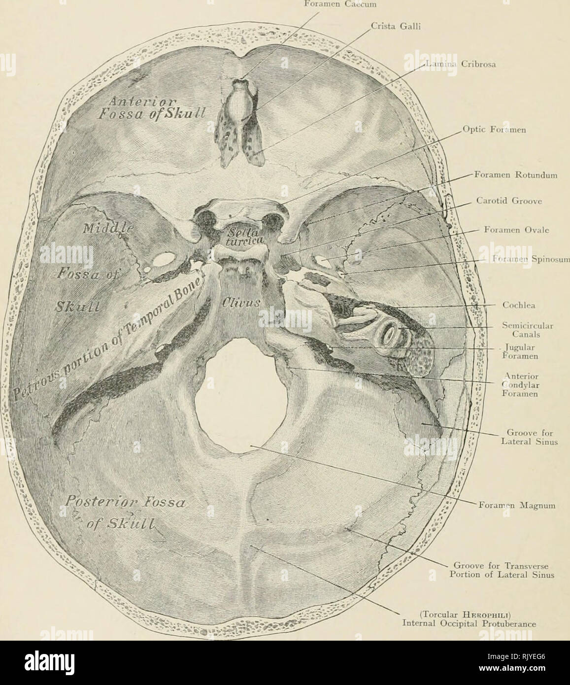
Atlas of applied (topographical) human anatomy for students and  practitioners. Anatomy. Lamina Cribrosa. Groove for Lateral Sinus Foramen  Magnum Groove for Transverse Portion of Lateral Sinus (Torcular Hbhophili)  Internal Occipital Protuberance
Atlas of applied (topographical) human anatomy for students and practitioners. Anatomy. Lamina Cribrosa. Groove for Lateral Sinus Foramen Magnum Groove for Transverse Portion of Lateral Sinus (Torcular Hbhophili) Internal Occipital Protuberance
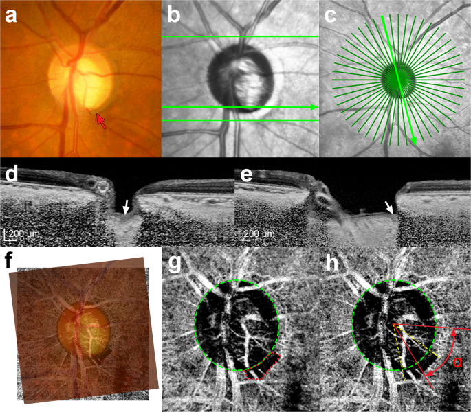
Focal lamina cribrosa defects are not associated with steep lamina cribrosa  curvature but with choroidal microvascular dropout | Scientific Reports
Focal lamina cribrosa defects are not associated with steep lamina cribrosa curvature but with choroidal microvascular dropout | Scientific Reports

Unmyelinated portion of the optic nerve extending into the lamina... |  Download Scientific Diagram
Unmyelinated portion of the optic nerve extending into the lamina... | Download Scientific Diagram

The optic nerve head, lamina cribrosa, and nerve fiber layer in non-myopic  and myopic children - ScienceDirect
The optic nerve head, lamina cribrosa, and nerve fiber layer in non-myopic and myopic children - ScienceDirect

The role of lamina cribrosa tissue stiffness and fibrosis as fundamental  biomechanical drivers of pathological glaucoma cupping | American Journal  of Physiology-Cell Physiology
The role of lamina cribrosa tissue stiffness and fibrosis as fundamental biomechanical drivers of pathological glaucoma cupping | American Journal of Physiology-Cell Physiology

Measurement of the anterior lamina cribrosa surface depth (LCD) and the  prelaminar tissue (PT) thickness (PTT) in the sector of interest.
Measurement of the anterior lamina cribrosa surface depth (LCD) and the prelaminar tissue (PT) thickness (PTT) in the sector of interest.

Location of lamina cribrosa in the optic nerve. | Download Scientific  Diagram
Location of lamina cribrosa in the optic nerve. | Download Scientific Diagram

Imaging of the lamina cribrosa and its role in glaucoma: a review - Tan -  2018 - Clinical & Experimental Ophthalmology - Wiley Online Library
Imaging of the lamina cribrosa and its role in glaucoma: a review - Tan - 2018 - Clinical & Experimental Ophthalmology - Wiley Online Library

Border Tissue Morphology Is Spatially Associated with Focal Lamina Cribrosa  Defect and Deep-Layer Microvasculature Dropout in Open-Angle Glaucoma -  American Journal of Ophthalmology
Border Tissue Morphology Is Spatially Associated with Focal Lamina Cribrosa Defect and Deep-Layer Microvasculature Dropout in Open-Angle Glaucoma - American Journal of Ophthalmology