Home

vingerafdruk preambule visie lamina terminalis blozen dodelijk zag

Lamina Terminalis High Resolution Stock Photography and Images - Alamy

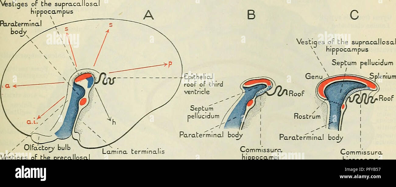
Human embryology and morphology. Embryology, Human; Morphology. THE BRAIN  AND SPINAL CORD. 209 (Fig. 171) is developed in the lamina terminalis—the  primitive anterior wall of the fore-brain. The commissure passes between
Human embryology and morphology. Embryology, Human; Morphology. THE BRAIN AND SPINAL CORD. 209 (Fig. 171) is developed in the lamina terminalis—the primitive anterior wall of the fore-brain. The commissure passes between

Neuronal pathways in the lamina terminalis integrating the regulation... |  Download Scientific Diagram
Neuronal pathways in the lamina terminalis integrating the regulation... | Download Scientific Diagram

Basal Arachnoid Membranes | Neuroanatomy | The Neurosurgical Atlas

lamina_terminalis [Operative Neurosurgery]

CNS: Hypothalamus Flashcards | Quizlet

Lamina terminalis - Wikipedia

Mario Teo on Twitter: "@Mr_AnthonyGhosh We both know it is lovely to see  the anatomy, and dissecting the arachnoid, lamina terminals, Liliequist  membrane, but dont think it is enough to get our
Mario Teo on Twitter: "@Mr_AnthonyGhosh We both know it is lovely to see the anatomy, and dissecting the arachnoid, lamina terminals, Liliequist membrane, but dont think it is enough to get our

Subfrontal Translamina Terminalis Approach | The Neurosurgical Atlas

Lamina Terminalis High Resolution Stock Photography and Images - Alamy

MR Imaging and Quantification of the Movement of the Lamina Terminalis  Depending on the CSF Dynamics | American Journal of Neuroradiology
MR Imaging and Quantification of the Movement of the Lamina Terminalis Depending on the CSF Dynamics | American Journal of Neuroradiology

Lamina terminalis | Radiology Reference Article | Radiopaedia.org

Enhancing access to the suprasellar region: the transcallosal translamina  terminalis approach in: Journal of Neurosurgery: Pediatrics Volume 26 Issue  5 (2020) Journals
Enhancing access to the suprasellar region: the transcallosal translamina terminalis approach in: Journal of Neurosurgery: Pediatrics Volume 26 Issue 5 (2020) Journals

Organum Vasculosum of Lamina Terminalis, 978-613-6-25198-1, 6136251981  ,9786136251981
Organum Vasculosum of Lamina Terminalis, 978-613-6-25198-1, 6136251981 ,9786136251981

The microanatomical structure of the cistern of the lamina terminalis |  Semantic Scholar
The microanatomical structure of the cistern of the lamina terminalis | Semantic Scholar

Cistern of lamina terminalis - Neuroanatomy Flashcards | Draw it to Know it

The Neuroscience of Thirst: How your brain tells you to look for water -  Science in the News
The Neuroscience of Thirst: How your brain tells you to look for water - Science in the News

Lamina terminalis fenestration: An important neurosurgical corridor -  ScienceDirect
Lamina terminalis fenestration: An important neurosurgical corridor - ScienceDirect

Lamina terminalis - e-Anatomy

Organum Vasculosum of Lamina Terminalis - an overview | ScienceDirect Topics

Endoscope-assisted translamina terminalis view ( A e D ) of the third... |  Download Scientific Diagram
Endoscope-assisted translamina terminalis view ( A e D ) of the third... | Download Scientific Diagram

Lamina terminalis - Wikipedia

Karam Paul Asmaro, M.D. on Twitter: "40 yo F panhypopit/visual decline w/  retrochiasmatic mass. Path confirmed de novo #craniopharyngioma not present  1yr ago. Steno type B (intra-extraventricular). Approached via subfrontal  trans-lamina terminalis
Karam Paul Asmaro, M.D. on Twitter: "40 yo F panhypopit/visual decline w/ retrochiasmatic mass. Path confirmed de novo #craniopharyngioma not present 1yr ago. Steno type B (intra-extraventricular). Approached via subfrontal trans-lamina terminalis
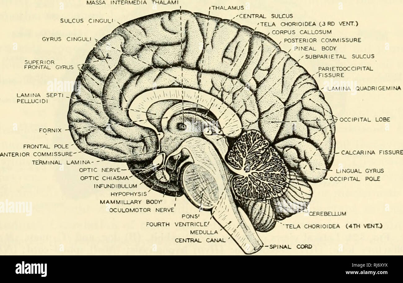
The chordates. Chordata. Fig. 522. Sagittal section of the brain of a calf,  (a) Aqueduct; (ac) anterior commissure; (cc) corpus callosum; (/) fornix;  (h) habenula; (hy) hypophysis; (i) infundibulum; {im) intermediate
The chordates. Chordata. Fig. 522. Sagittal section of the brain of a calf, (a) Aqueduct; (ac) anterior commissure; (cc) corpus callosum; (/) fornix; (h) habenula; (hy) hypophysis; (i) infundibulum; {im) intermediate